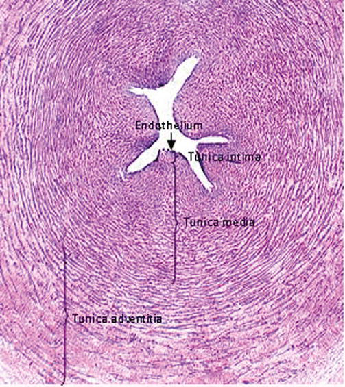
Fetal Pig Anatomy I External Features, Oral Cavity, Pharynx, and Digestive System Umbilical Artery Umbilical Vein 13 Post Laboratory Questions (Day 2) Name ___ _____ _ 1 Which type of vessels, arteries or veins, has more muscle fibers?Gross anatomy Origin The umbilical artery originates from the anterior division of the internal iliac artery In the fetus, it travels within the umbilical cord to the placenta and hence is the communication to the maternal circulation After the umbilical cord has been cut at birth, clots form within the vessel and it obliteratesQualitative and quantitative changes in the human umbilical artery and vein were observed in 15 human specimens at different stages of development Features such as intimal thickening and cellular lipid accumulation were found in umbilical vasculature Cellular origin
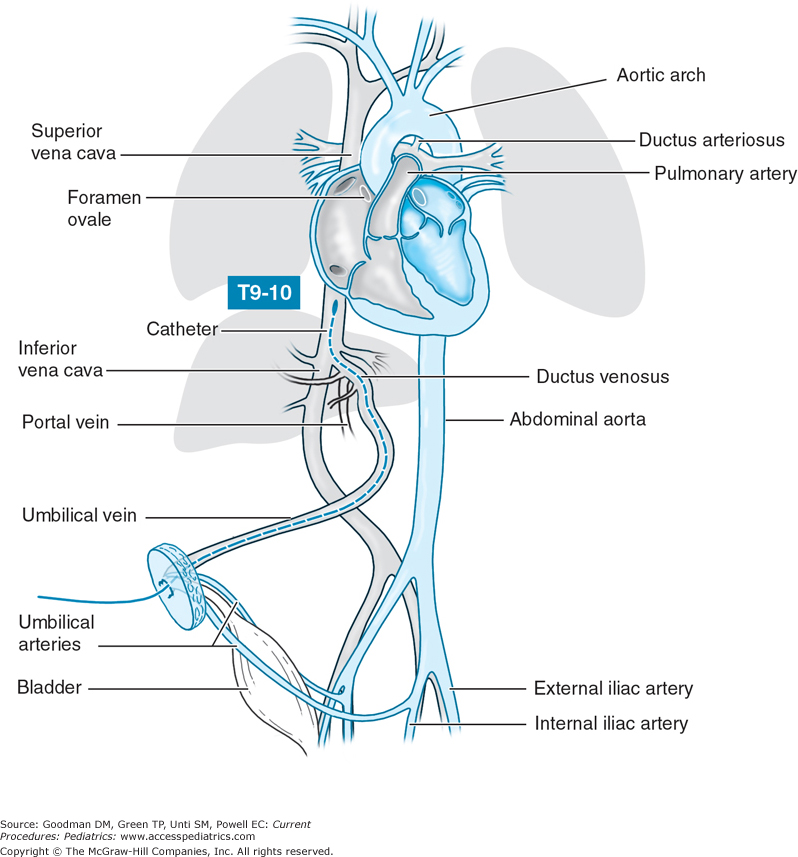
Umbilical Vein Catheter Indications And Procedure Medcaretips Com
Umbilical artery vein anatomy
Umbilical artery vein anatomy-Umbilical arteries in the cord carry rich carbondioxide (deoxygenated) and waste products from the baby to the placenta The placenta gets rid of these givinUmbilical cord artery and vein There are usually two umbilical arteries present together with one umbilical vein in the umbilical cord Single umbilical artery fetuses and neonates had a 6 The umbilical cord also being called the 'navel string' or 'birth cord,' is a channel between the developing fetus and the placenta In the placenta, the arteries distribute the blood to reach the


Article On Umbilical Cord Standard Of Care
De très nombreux exemples de phrases traduites contenant "umbilical vein blood" – Dictionnaire françaisanglais et moteur de recherche de traductions françaisesWhat is the functional significance of this?PDF On , Aymelek Çetin published ULTRASTRUCTURE OF HUMAN UMBILICAL ARTERY AND VEIN Find, read and cite all the research you need on ResearchGate
From the heart's right ventricle, deoxygenated blood travels to the lungs via the pulmonary artery With the help of capillaries, the blood oxygenates through the respiration process and is returned to the heart via the pulmonary veins In pregnancy, the artery passes deoxygenated blood to the placenta, and the umbilical vein inside the cord carries oxygenatedUmbilical vein varix (UVV) is a rare, idiopathic, focal dilatation of the umbilical vein, either within the intraamniotic portion of the umbilical cord or within the fetal abdomen On ultrasound (US), UVV appears as a round or fusiform anechoic structure that is located either within the umbilical cord or within the fetal abdomen, inferior to the fetal liver and close to the anterior abdominalOccasionally, there is only the one single umbilical artery (SUA) present in the umbilical cord This is sometimes also called a twovessel umbilical cord, or twovessel cord Approximately, this affects between 1 in 100 and 1 in 500 pregnancies, making it the most common umbilical abnormality Its cause is not known
Just ask here or over at Template talkInfobox anatomyUmbilical vein marsupialization has been described as a onestep or twostep procedure In the onestep procedure, a ventral midline celiotomy is performed and the infected umbilical vein isolated The umbilicus is excised and the umbilical vein sutured closed A sterile glove may be placed over the stump to prevent abdominal contamination Next, the umbilical vein isThe umbilical artery is a paired artery (with one for each half of the body) that is found in the abdominal and pelvic regions In the fetus, it extends into the umbilical cord


Emergent Umbilical Venous Catheter Uvc Placement Brown Emergency Medicine


Umbilical Artery Catheter Indications And Procedure Medcaretips Com
A firm understanding of the anatomy of the umbilical vein and its further course is crucial for correct catheter placement Umbilical vein catheters may be placed in the inferior vena cava above the level of the ductus venosus and below the level of the right atrium (1012 cm) This acts as central venous access, allowing central venous pressure (CVP) monitoring, medicationThe umbilical cord contains _____ umbilical vein (s) and _____ umbilical artery (ies) asked in Anatomy & Physiology by Mandi A2 In general, we have no conscious control over smooth muscle or cardiac muscle function, whereas
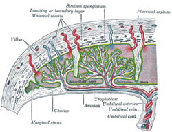


Umbilical Artery Wikipedia


Histology Of Umbilical Cord In Mammals Intechopen
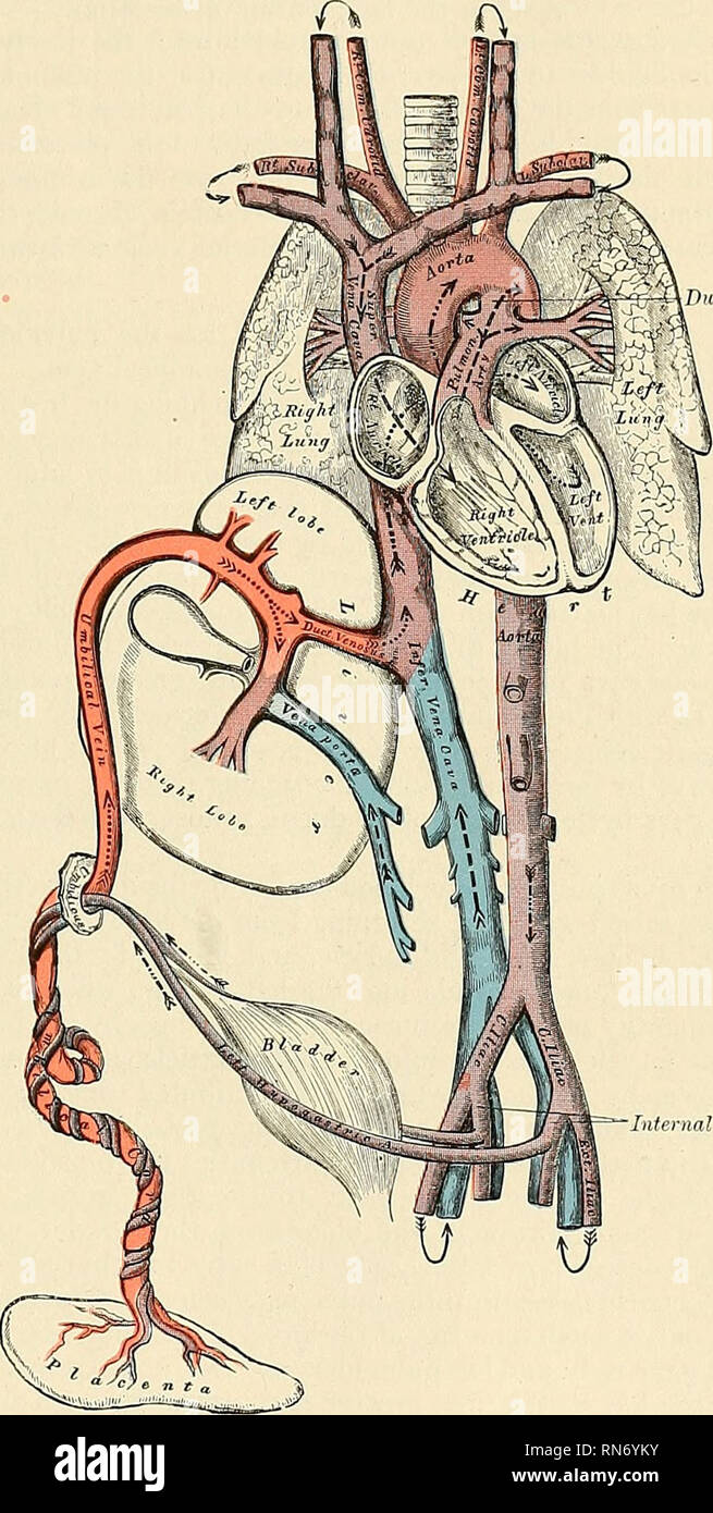
Umbilical Artery and Vein Diagram with Compression Medical Illustration, Human Anatomy Drawing This image may only be used in support of a single legal proceeding and for no other purpose Read our License Agreement for details To license thisHenry Gray (15–1861) Anatomy of the Human Body 1918 the umbilical opening, the two arteries, now termed umbilical, enter the umbilical cord, where they are coiled around the umbilical vein, and ultimately ramify in the placentaTherefore, the umbilical venous catheter and the umbilical artery catheter can be easily distinguished on abdominal radiographs The —Postmortem venogram in 27week premature neonate shows normal anatomy Umbilical vein enters left portal vein opposite origin of ductus venosus Focal dilatation of umbilical vein just inferior to its insertion into left portal vein is umbilical


Umbilical Cord Artery And Vein Link Pico


Maternal Artery Points Maternal Vein Umbilical Cord Chegg Com
The umbilical cord contains _____ umbilical vein(s) and _____ umbilical artery(ies) asked in Anatomy & Physiology by Steph A two;The umbilical artery was the first vessel studied by obstetric Doppler examination Flow velocity waveforms from the umbilical artery represent the downstream or placental resistance to flow Umbilical artery resistance decreases progressively throughout gestation, reflecting the increase and dilation in villous vascularizationThis template is a customized wrapper for {{Infobox anatomy}}Only some fields from {{Infobox anatomy}} can work, which you can see on the documentation page for each infobox Questions?


Dams Nepal Fetalcirculation The Fetal Prenatal Circulation Differs From Normal Postnatal Circulation Mainly Because The Lungs Are Not In Use Instead The Fetus Obtains Oxygen And Nutrients From The Mother Through


1
The umbilical arteries are the direct continuation of the internal iliac arteries A catheter passed into an umbilical artery will usually (but not always) enter the aorta via the internal iliac artery Its path is, therefore, initially inferior and lateral as it passes around the bladder, before turning cephalad and medial to enter the aortaThe portion of the vessel gets replaced by fibrous tissue due to the lack of blood flow in the distal part of the umbilical artery This ligament is also referred to as the cord of the umbilical artery Misplacement can occur amongst other places, in normal anatomy, at the level of the left portal vein and at the level of the hepatic veins The umbilical vein closes soon after birth (7 days) and becomes the round ligament of the liver This lies in the free edge of the falciform ligament and is also continuous with the ligamentum venosum (the remnant of the ductus venosus) An umbilical
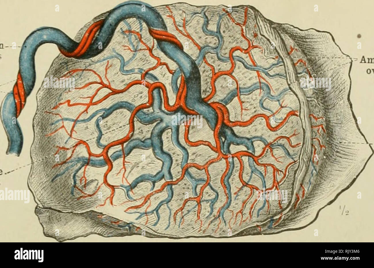


Page 2 Umbilical Vein High Resolution Stock Photography And Images Alamy


Umbilical Cord Of The Amazonian Manatee A Cross Section Showing Two Download Scientific Diagram
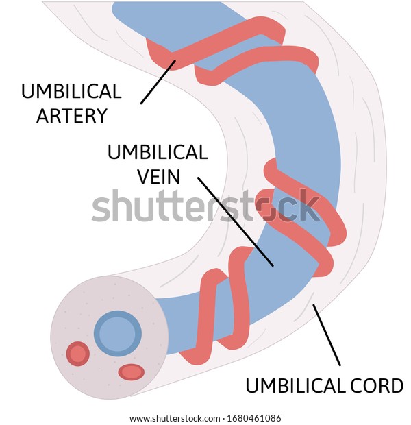
During the prenatal period, the umbilical artery is the main continuation of the internal iliac artery The two umbilical arteries run through the umbilical cord, comprising a helix around the umbilical vein The arteries carry deoxygenated and nutrientdeficient blood from the fetus to the placenta After birth, when the umbilical cord is cut, a blood clot forms and occupies the distal portion of the artery In the following months, the distal part of the umbilical arteryQualitative and quantitative changes in the human umbilical artery and vein were observed in 15 human specimens at different stages of development Features such as intimal thickening and cellular lipid accumulation were found in umbilical vasculature Cellular origin and quantification of lipidcontaining cells were determined by electron microscopy The medial umbilical ligament is the distal obliterated portion of the umbilical artery It develops after birth when the umbilical cord is cut;


A Radiologist S Guide To The Performance And Interpretation Of Obstetric Doppler Us Radiographics


Umbilical Artery Wikipedia
Normal umbilical cord anatomy consists of three vessels represented by two umbilical arteries and one umbilical vein By the seventh week of gestation, the right umbilical vein usually obliterates, leaving a single (left) umbilical vein patent However, there have been documented cases of umbilical cords containing fourvessels The persistence of two umbilical veins and two umbilicalPelvis Artery And Vein Innervation Diagram In this image, you will find inferior mesenteric artery, lumbar artery, ureter, ovarian artery, iliolumbar artery, middle sacral artery, superior rectal artery, internal iliac artery, external iliac artery in it You may also find umbilical artery, obturator artery, accessory obturator arteryAnatomy_umbilical_vein 1/2 Anatomy Umbilical Vein eBooks Anatomy Umbilical Vein Vascular Biology of the PlacentaYuping Wang The placenta is an organ that connects the developing fetus to the uterine wall, thereby allowing nutrient uptake, waste elimination, and gas exchange via the mother's blood supply Proper vascular development in the placenta is


1


Fetal Circulation Wikipedia
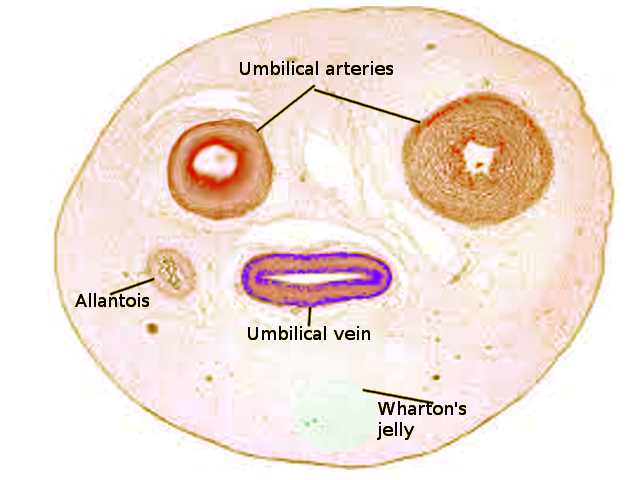
Single Umbilical Artery Axial US shows only 2 vessels in the umbilical cord The umbilical artery (UA) has a thicker wall than the umbilical vein (UV) The increased diameter of the single umbilical artery (SUA) is most apparent on crosssectional views Gross image and longitudinal US of an umbilical cord with a SUA show a hypocoiled cord Abnormalities in the number of vessels can be found for both the umbilical artery and vein We sometimes encounter cases of a decreased number of umbilical cord vessels, such as a single umbilical artery In contrast, there may be an increase from three to four vessels within the umbilical cord A supernumerary umbilical vein is particularly very rare, and it is generally foundThe umbilical vein is the large, red vessel at the far left The umbilical arteries are purple and wrap around the umbilical vein Scheme of placental circulation The umbilical artery is a paired artery (with one for each half of the body) that is found in the abdominal and pelvic regions


Anatomy Of Umbilical Cord Two Umbilical Veins And One Umbilical Artery Royalty Free Cliparts Vectors And Stock Illustration Image


Fetal Circulation Quiz Maternity Nursing Nclex
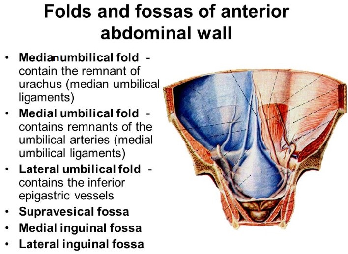
The medial umbilical ligament (or cord of umbilical artery, or obliterated umbilical artery) is a paired structure found in human anatomy It is on the deep surface of the anterior abdominal wall, and is covered by the medial umbilical folds (plicae umbilicales mediales)The umbilical vein is a vein present during fetal development that carries oxygenated blood from the placenta into the growing fetus The umbilical vein provides convenient access to the central circulation of a neonate for restoration of blood volume and for administration of glucose and drugs It is enclosed inside a tubular sheath of amnion and consists of two paired umbilical arteries and one umbilical vein During development, the umbilical arteries have a vital function of carrying deoxygenated blood away from the fetus to the placenta However, after birth, a significant distal portion of the umbilical artery degenerates


Umbilical Arteries An Overview Sciencedirect Topics


Single Umbilical Artery Wikipedia
The umbilical vein arises from multiple small veins within the placenta which carry oxygen and nutrient rich blood derived from the maternal blood circulation via the chorionic villi From here, it enters the umbilical cord, along with the paired umbilical arteries0 Answers 0 votes answered by illeztg Best answer Answer D 0 votes answered by nnorment Thank you!The Inferior Epigastric artery also brings about anterior pubic artery, which associated with the Iliopubic vein crosses the superior pubic ramus In 2530% (some studies mention as high as 7080%) of people, the anterior pubic branch is large and can replace the obturator artery (Obturator artery originating from the Hypogastric artery is missing) This large arterial branch (Aberrant
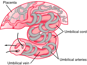


Umbilical Definition Of Umbilical By Medical Dictionary


Single Umbilical Artery Anatomy Of Umbilical Cord With One Umbilical Artery And One Umbilical Vien Stock Illustration Download Image Now Istock
Umbilical cord 1 Vein and 1 artery a alexistepp at 1257 PM Dr called and said looking at week ultrasound baby only has one vein and one artery She said that this could indicate she has a genetic disorder, such as Down syndrome, autism etc she suggested to get the amino fluid test but I have heard there's a lot of risks These are not branches but are actually a direct continuation of the umbilical artery Umbilical cord anatomy At birth the umbilical cord which provided vascular flow between the fetus and placenta is clamped and cut The umbilical cord is made up of a substance called whartons jelly instead of normal connective tissue and skin The umbilical vein goes all the wayInsertion Of Umbilical Lines (UAC, UVC) Neonatal Clinical Guideline V30 Page 8 of 16 Note arterial line dips into iliac Fig1 4 (Drawn from postmortem venogram image by J Clegg, RCH) Umbilical vein anatomy TO CALCULATE INSERTION LENGTH UAC = 3 UAC T6 x weight 9 stump UVC = 15 x weight 55 stump Use in emergency length ~ 5cm


Lesions Of The Umbilical Cord In Newborn Pedchrome Pediatrician Blog On Newborn Child Health


A Partially Dissected Human Uc Showing Umbilical Arteries Vein Download Scientific Diagram
The umbilical artery (latin arteria umbilicalis) is a branch of the anterior division of the internal iliac artery that functions only in the time of the placental blood circulation The umbilical artery passes along the anterior wall of the abdominal cavity to the umbilical ring, it passes further in the umbilical cord to the placentaThe umbilical artery is a paired artery that is found in the abdominal and pelvic regions In the fetus, it extends into the umbilical cordThis video is tarTHE umbilical vein has been supposed to undergo thrombosis and fibrosis in the postnatal period 1,2 Despite this, it has been shown that the adult umbilical vein can be cannulated in a superficial position in the upper abdomen, and through it direct access to the portal venous system can be obtained 3 If the umbilical vein had become fibrosed and obliterated in infancy, it should not be
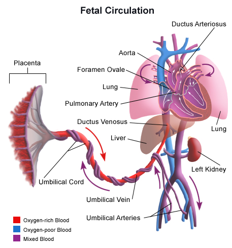


Fetal Circulation


How To Perform Umbilical Cord Arterial And Venous Blood Sampling In Neonatal Foals Sciencedirect
ANATOMY The umbilicus is a remnant of the fetalmaternal connection At birth the structure consists of the paired umbilical arteries, a single umbilical vein, and the urachus Before birth, the umbilical vein serves as the source of oxygenated blood to the fetus via the liver and ductus venosus/portal veinGross anatomy The portal vein usually measures approximately 8 cm in length in adults with a maximum diameter of 13 mm It originates posterior to the neck of the pancreas where it is classically formed by the union of the superior mesenteric and splenic veins (the portovenous confluence) The origin of the vein defines the location of the pancreatic neckCorresponding Author Aymelek Cetin, Inonu University Faculty of Medicine, Department of Anatomy, Malatya, Turkey Email aymelekcetin@inonuedutr 6 INTRODUCTION The umbilical cord contains two arteries and one vein (1,2) The lumen of umbilical vein is large and has an irregular shape Regarding the umbilical artery wall, this is greater
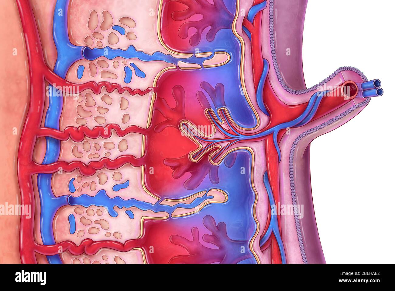


Umbilical Artery High Resolution Stock Photography And Images Alamy
.jpg)


Fetal Circulation And Erythropoiesis Embryology Medbullets Step 1
The umbilical vein is the conduit for blood returning from the placenta to the fetus until it involutes soon after birth The umbilical vein arises from multiple tributaries within the placenta and enters the umbilical cord, along with the (usually) paired umbilical arteries Once it enters the fetus at the umbilicus, it courses upwards towards theUmbilical vein to artery ratio in fetuses with single umbilical artery Sepulveda W(1), Peek MJ, Hassan J, Hollingsworth J Author information (1)Centre for Fetal Care, Royal Postgraduate Medical School, Queen Charlotte's and Chelsea Hospital, London, UK Comment in Ultrasound Obstet Gynecol 1996 Jul;8(1)57 In fetuses with single umbilical artery (SUA) the entire blood flow toUmbilical arterial (UA) Doppler assessment is used in surveillance of fetal wellbeing in the third trimester of pregnancy Abnormal umbilical artery Doppler is a marker of placental insufficiency and consequent intrauterine growth restriction (IUGR) or suspected preeclampsia Umbilical artery Doppler assessment has been shown to reduce perinatal mortality and morbidity in high
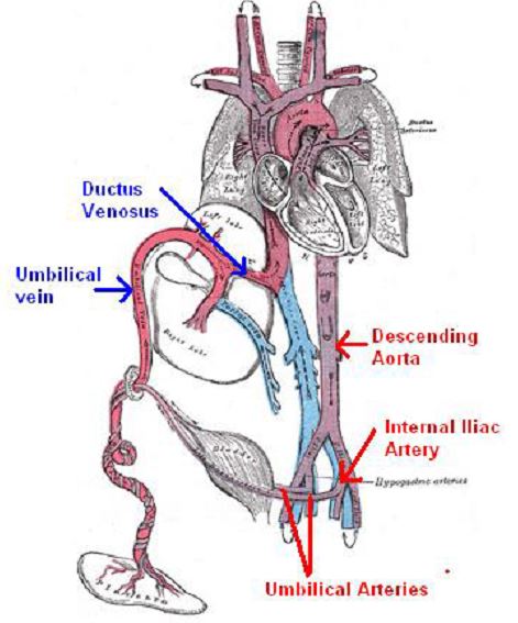


Umbilical Catheters
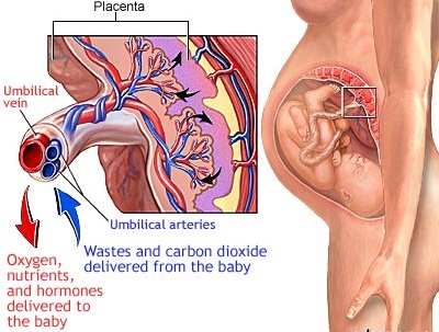


In The Human Umbilical Cord Are There 2 Arteries And 1 Vein Or 2 Veins And 1 Artery Socratic


How Many Arteries And Veins Does The Umbilical Cord Contain Anatomy Valuemd Usmle Qbank


Comprehensive Imaging Review Of Abnormalities Of The Umbilical Cord Radiographics


Umbilical Cord Blood Gases And Birth Asphyxia


Umbilical Cord Radiology Key
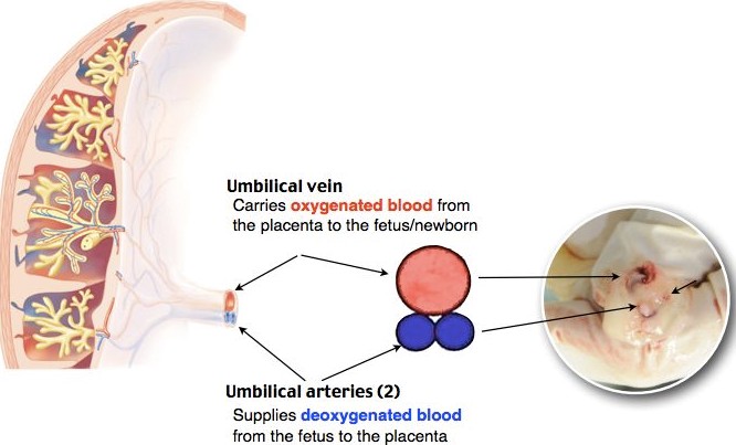


Umbilical Artery Umbilical Vein 네이버 블로그
:background_color(FFFFFF):format(jpeg)/images/library/13725/Umbilical_vein.png)


Umbilical Vein Anatomy Tributaries Drainage Kenhub


1 Label The Maternal Artery And Vein A Why Is The Chegg Com
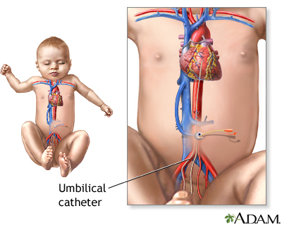


Umbilical Catheters Information Mount Sinai New York
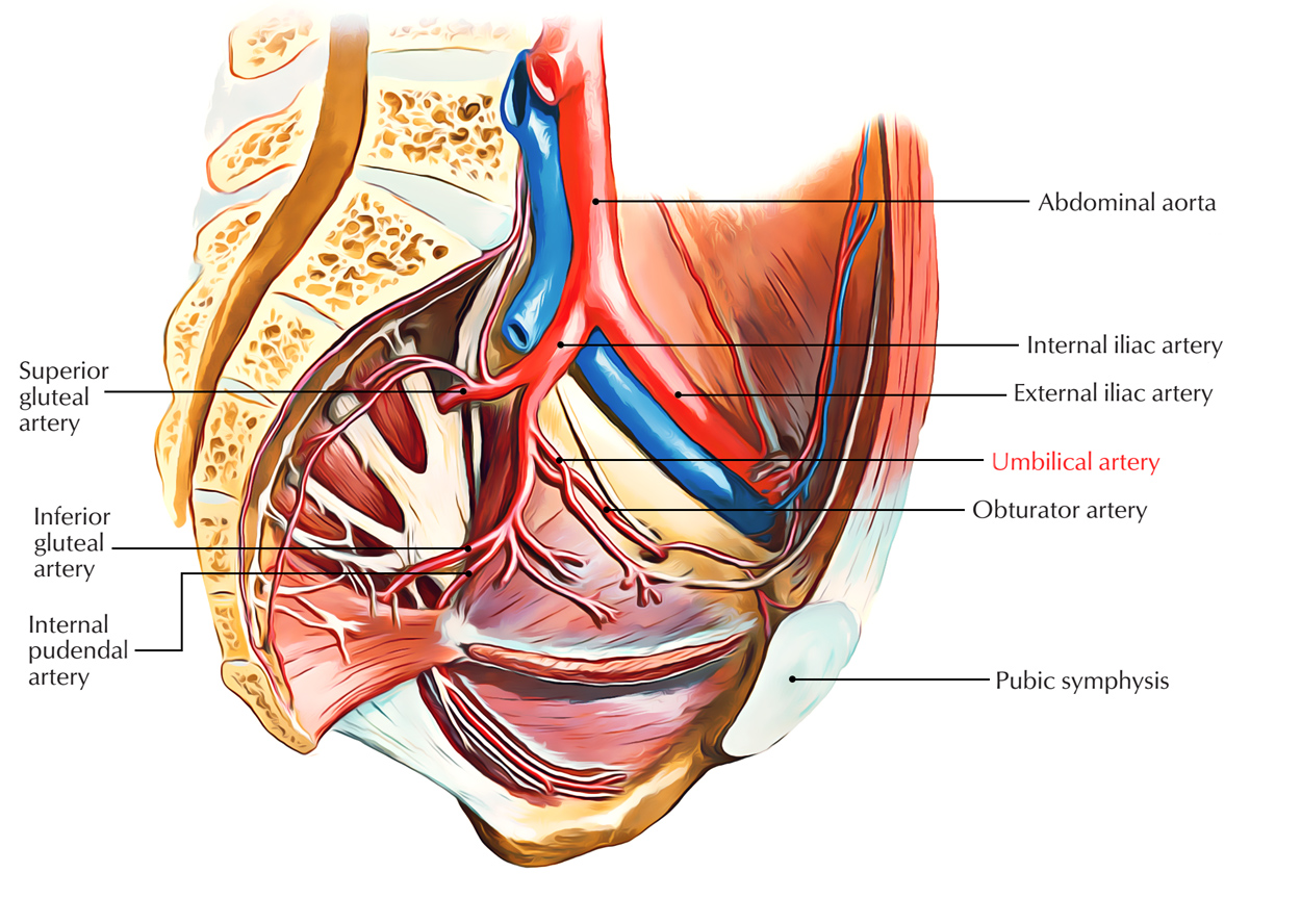


Umbilical Artery Earth S Lab


Umbilical Vein Catheterization 60 Second Em
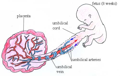


What Type Of Blood Do Each Of The Umbilical Blood Vessels Carry Socratic
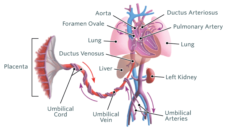


Fetal Blood And Circulation Changes Of Fetal Circulation After Birth In The Newborn Science Online


Placenta Umbilical Cord Stock Illustrations 998 Placenta Umbilical Cord Stock Illustrations Vectors Clipart Dreamstime
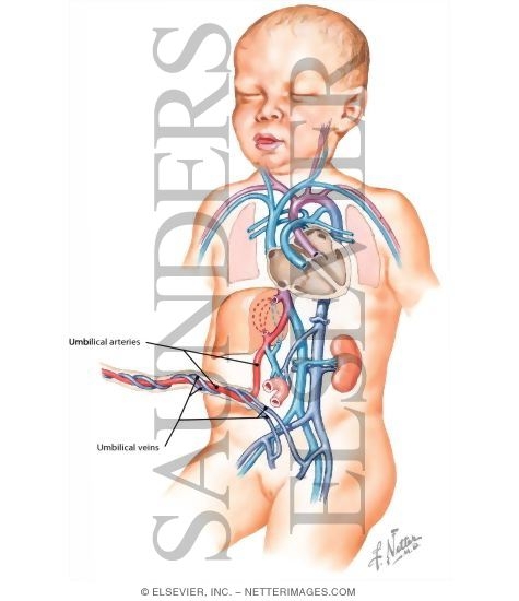


Umbilical Cord
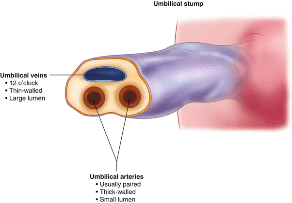


Umbilical Venous Catheters Insertion And Removal Springerlink


What Is The Umbilical Cord Definition Function Video Lesson Transcript Study Com


Jaypeedigital Ebook Reader


Amazon Com Jin Human Anatomical Placenta Model With Digital Identification Umbilical Cord Model Medical School Teaching Tools Industrial Scientific


Embryology Flashcards Quizlet
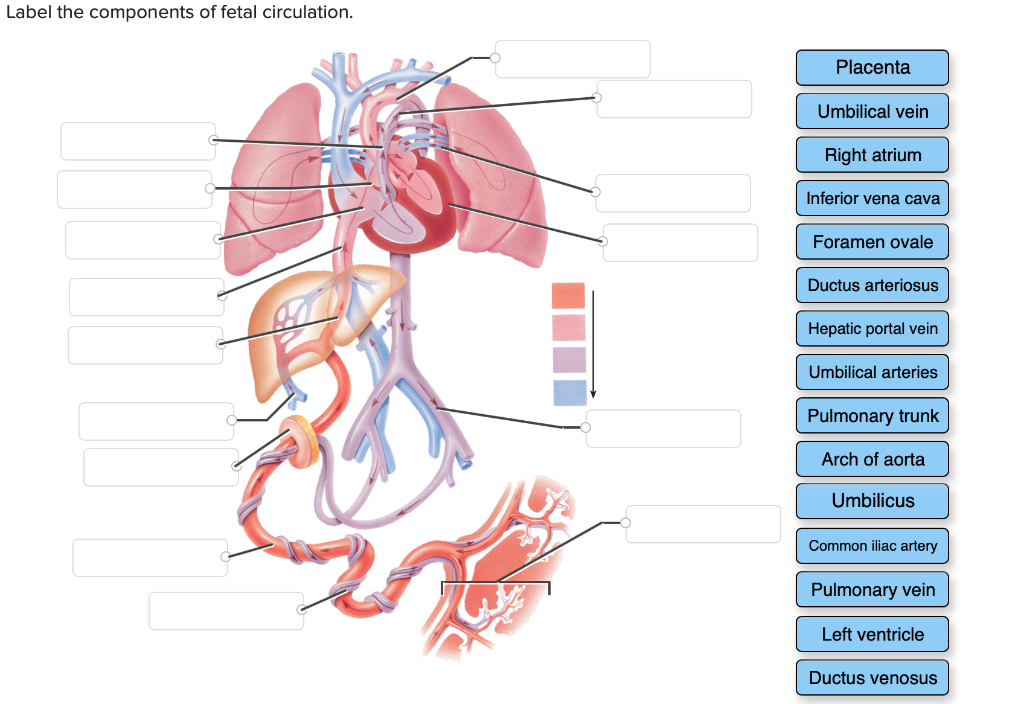


Label The Components Of Fetal Circulation Placenta Chegg Com


Umbilical A In Foetus Human Body Anatomy Arteries Anatomy Medical Anatomy


Umbilical Cord Structure Functions Storage Abnormalities Infections


Schematic Representation Of Structure Of Umbilical Cord In Cattle Download Scientific Diagram


1


Umbilical Vein Catheter Indications And Procedure Medcaretips Com


Structure Of Umbilical Cord Blood Schematic Image Download Scientific Diagram


Miscellaneous Anatomy And Embryology Flashcards Quizlet


Umbilical Artery Catheterization Obgyn Key
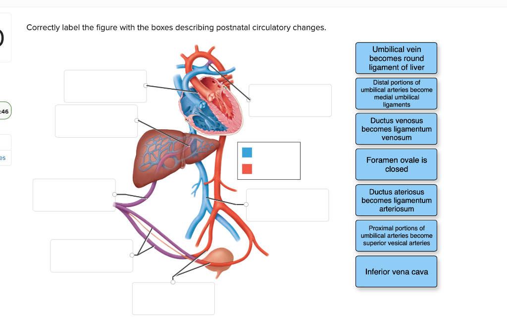


Label The Components Of Fetal Circulation Placenta Chegg Com


Article On Umbilical Cord Standard Of Care


Abdominal Aortic Perforation By An Umbilical Arterial Catheter Adc Fetal Neonatal Edition


Fetal Circulation Wikiradiography
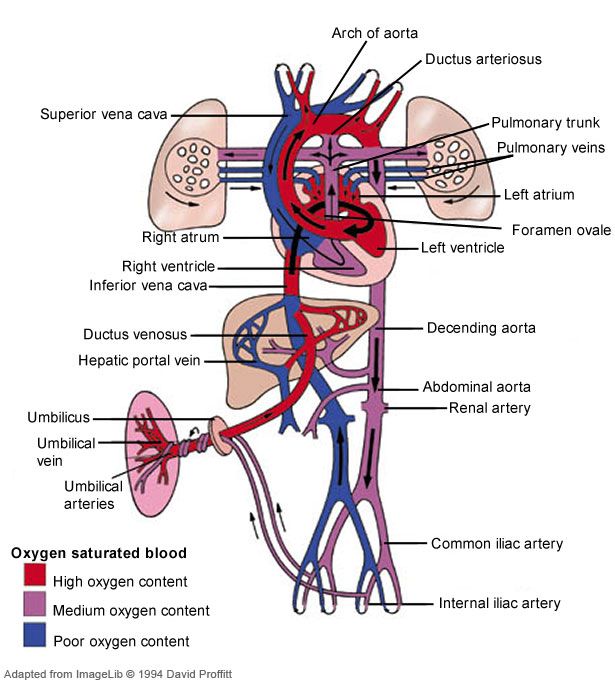


What Type Of Blood Do Each Of The Umbilical Blood Vessels Carry Socratic
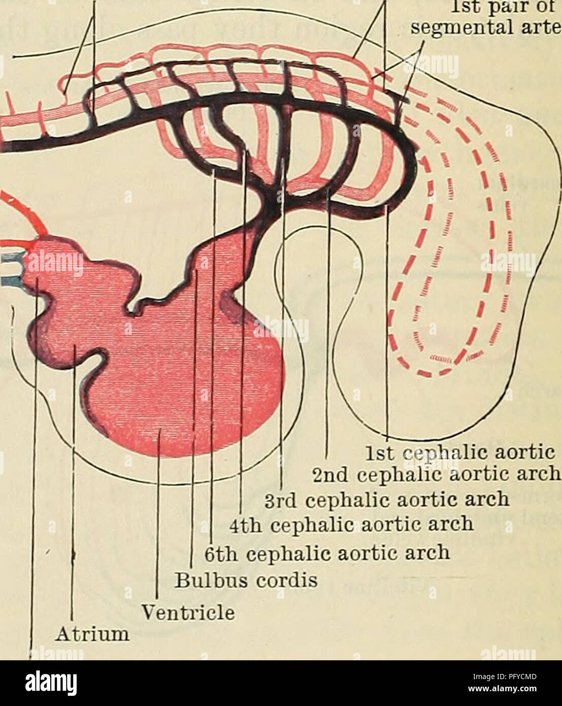


Umbilical Anatomy Anatomy Drawing Diagram
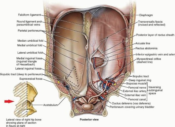


Umbilical Artery Umbilical Vein 네이버 블로그
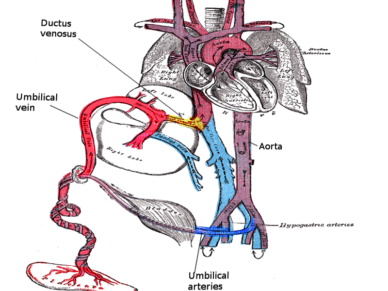


Umbilical Vein Catheterization Article


Umbilical Vein An Overview Sciencedirect Topics
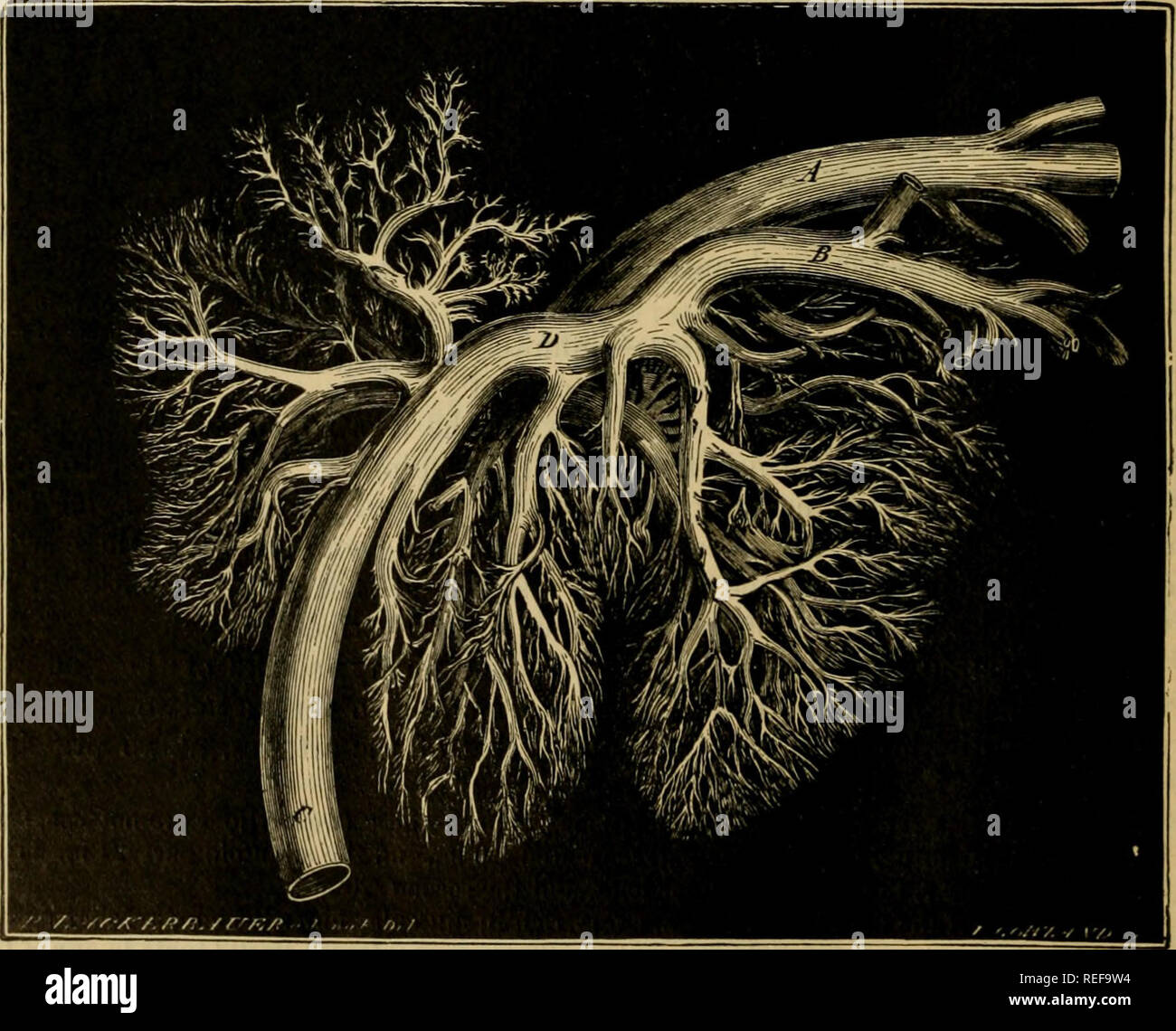


The Comparative Anatomy Of The Domesticated Animals Veterinary Anatomy Ftetus Opened On The Left Side To Show The Course Of The Umbiucal Vessei S In The Body A Umbilical Cord B Umbilical


Histology Of The Umbilical Cord Youtube
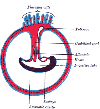


Anatomy Abdomen And Pelvis Umbilical Cord Article


The Schematic Cross Section Of Human Umbilical Cord Covered With The Download Scientific Diagram
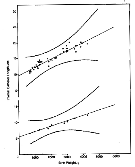


Umbilical Artery And Vein Catheterisation In The Neonate


Umbilical Artery Radiology Reference Article Radiopaedia Org


Umbilical Cord Radiology Key


Fetal Circulation Dr Najeeb Flashcards Quizlet
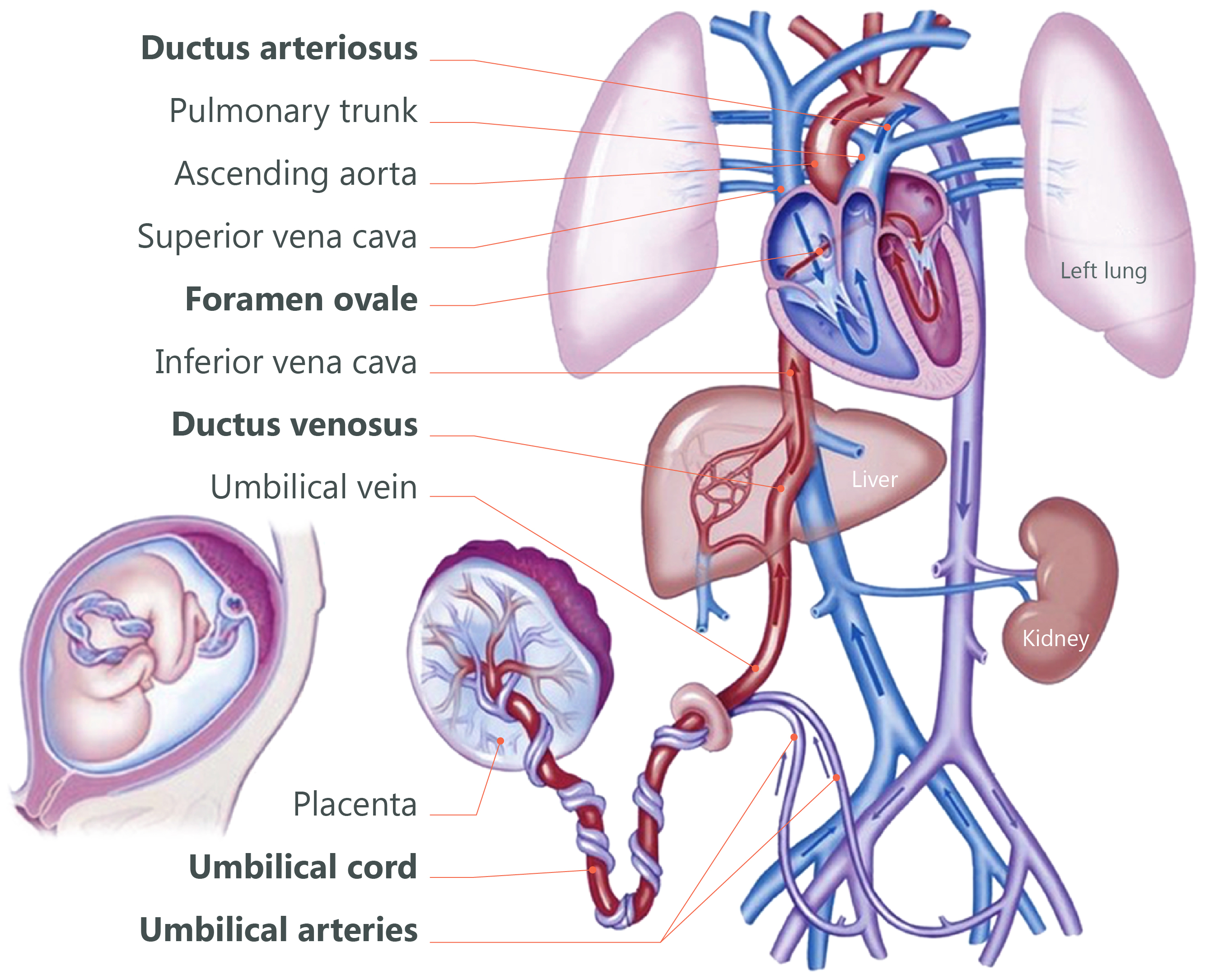


Cord Clamping Concord Neonatal


Figure Fetal Circulation Contributed By T Silappathikaram Statpearls Ncbi Bookshelf


Umbilical Vessels


Umbilical Vein Wikipedia


Cardiovascular System Blood Vessels Anatomy Chap Ppt Download


How To Perform Umbilical Cord Arterial And Venous Blood Sampling In Neonatal Foals Sciencedirect


Umbilical Vessel Catheterization Basicmedical Key
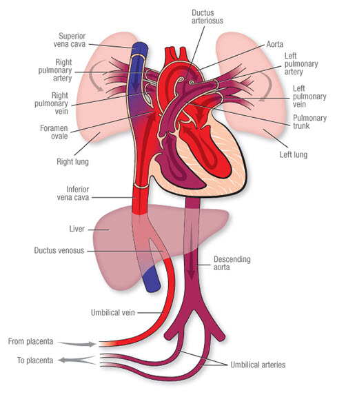


Fetal Circulation American Heart Association


1


Umbilical Veins An Overview Sciencedirect Topics
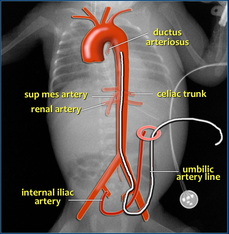


The Radiology Assistant Lines And Tubes In Neonates


Umbilical Cord Blood Gases And Birth Asphyxia


Histology Of Umbilical Cord In Mammals Intechopen


The Placenta Umbilical Cord And Amniotic Sac Knowledge Amboss
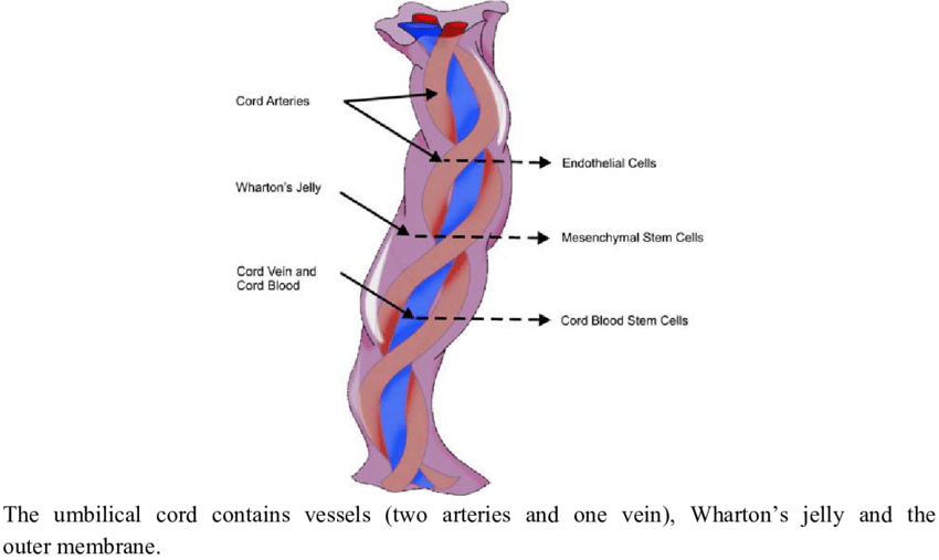


Umbilical Anatomy Anatomy Drawing Diagram


Umbilical Cord Normal Morphology Download Scientific Diagram


Anatomy Umbilical Cord Two Umbilical Arteries Stock Vector Royalty Free


Two Vessel Umbilical Cord What Does That Mean
:background_color(FFFFFF):format(jpeg)/images/article/en/umbilical-artery/a1dv25uodME8Io7y2dEpA_pgOFOniosFvo3tDaBiD9A_6umbilical_artery_magnified.png)


Umbilical Artery Anatomy Branches Supply Kenhub


Anatomy Descriptive And Applied Anatomy The Heart 569 The Peculiarities In The Arterial System Of The Fetus Are The Communication Between The Pulmonar Y Artery And The Descending Aorta By Means Of


Prenatal Diagnosis Of Type Ii Single Umbilical Artery Persistent Vitelline Artery In A Normal Fetus Gamez 13 Ultrasound In Obstetrics Amp Gynecology Wiley Online Library


The Umbilical Cord What It Is And How It Works Babycenter


Histology Of Umbilical Cord In Mammals Intechopen


Anatomy Abdomen And Pelvis Umbilical Cord Article


Anatomy Of Umbilical Cord Two Umbilical Veins And One Umbilical Artery Stock Illustration Illustration Of Newborn Blue


Umbilical Cord And Remnants Embryology Medbullets Step 1


Umbilical Artery


Umbilical Cord Wikipedia


Umbilical Cord Www Medicoapps Org


Umbilical Artery Umbilical Vein 네이버 블로그


How Is Umbilical Vein Catheterization Performed


0 件のコメント:
コメントを投稿